Tomography of the Atlas Vertebra
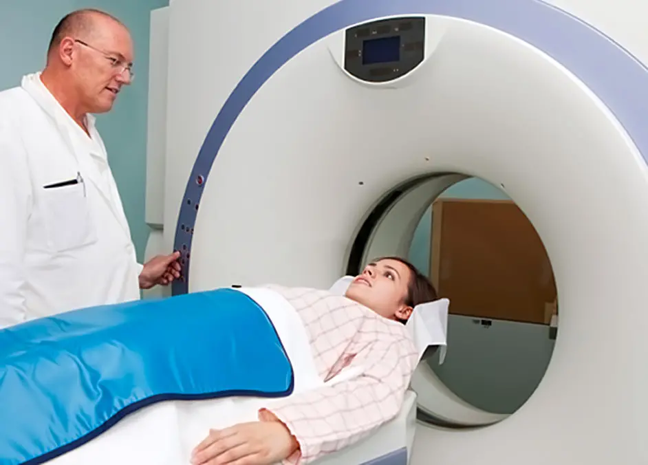
3D Spiral CT – Cone Beam CT – MRI
Since ancient times, humans have sought methods to observe the inside of the human body to understand its functioning and identify potential dysfunctions without resorting to invasive procedures. Today, technological advancements provide us with a wide range of diagnostic imaging tools, each with specific characteristics, limitations, advantages, and disadvantages. The choice of the most suitable diagnostic method depends on what needs to be visualized. Some techniques are better suited for certain types of analyses than others. As this is an important health-related topic, yet often confusing for those outside the field, we believe it is important to provide a clear and useful overview to navigate the various available options and avoid constantly confusing one exam with another.
Absence of Atlas Misalignment in Medical Reports
It may seem paradoxical, but expecting a doctor or radiologist to discuss Atlas misalignment is like assuming a house painter is also an expert on Picasso’s works. While both work in the field of painting, they operate in completely different domains. Sure, you might find a painter who, besides painting walls, is a passionate Picasso enthusiast, but in most cases, this is unlikely. This simple analogy aims to help you understand that a radiologist does not look for vertebral misalignments. This concept needs to be crystal clear.
Do not expect to find references to your Atlas misalignment in radiological reports. Only recently, and only after the development of a method and equipment capable of effectively and stably correcting the first cervical vertebra, has it been possible to retrospectively identify the Atlas as a potential cause of certain disorders. In medical literature, an Atlas misalignment is not yet considered a pathological condition, which is why doctors and radiologists generally do not conduct investigations in this direction. Consequently, even when Atlas misalignment is present and clearly visible, it is rarely identified or included in a radiological report.
Evolution of Technology and Specialized Expertise
If you want to explore and adopt new technology, it makes no sense to rely on someone who still uses outdated methods, regardless of their experience or training. For example, you cannot expect a gasoline car driver to provide reliable insights into what it’s like to drive an electric Tesla.
A Common Misconception About Medical Knowledge
It is a common misconception to think that a doctor must always be informed about every new development in medicine simply because they work in the healthcare sector. Medicine is constantly evolving, and your doctor may not necessarily be up-to-date on the latest discoveries, such as Atlas misalignment and its correction.
Relying on Specialists With Specific Expertise
Therefore, if you want reliable and in-depth information about the durable correction of the Atlas, you cannot and should not rely on your trusted doctor or therapist, as they are unlikely to have specific experience in this field. Their knowledge may be limited or inaccurate. Understanding this reality is essential for making informed decisions about how to proceed and whose expertise to trust.
The universal rule, valid in any field, is that when it comes to something revolutionary and unprecedented, only its creator has a complete understanding of what it is and how it truly works. Subsequently, this knowledge is gradually passed on to those within their circle. Everyone else, even if they operate in the same sector, remains excluded and can only speculate or hypothesize, often inaccurately or incorrectly.
In this context, Atlantomed stands as a reference point for effectively addressing upper cervical misalignment. If you want to achieve results that only Atlantomed can provide, it is essential to consult a professional who practices the Atlantomed method.
Diagnostic Methods Through Imaging
X-ray
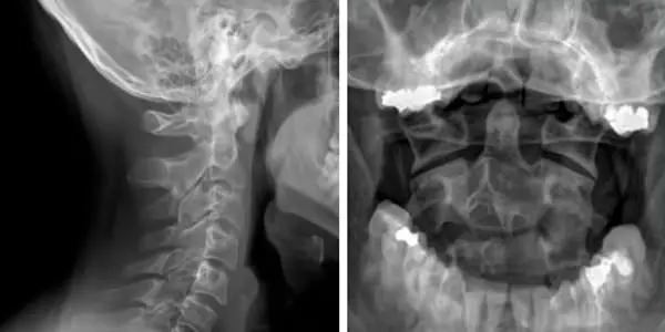
The X-ray (RX) was the first imaging method developed to examine the inside of the human body. It works as follows: an X-ray tube emits radiation that passes through the body and is then captured by a sensor positioned on the opposite side.
Due to their higher density, bones absorb more X-rays than soft tissues. This causes bones to appear white on the X-ray image, as they block more rays. In contrast, soft tissues, which allow more X-rays to pass through, appear darker on the final image.
What are X-rays? They are electromagnetic waves, similar to sunlight but with a much higher frequency. The higher the frequency, the greater the penetrating power of the electromagnetic waves. While sunlight can be blocked by a simple sheet of paper or the clothes we wear, X-rays can pass through the body, allowing its internal structures to be visualized.
Thanks to technological advancements and increased sensor sensitivity, very low doses of X-rays are now sufficient to produce a clear image.
An X-ray can only depict a subject in two dimensions. Therefore, an X-ray image cannot be used to evaluate the three-dimensional spatial relationship between the Atlas and the skull bone. Attempting to analyze Atlas misalignment in three spatial planes based on a two-dimensional X-ray demonstrates a lack of basic knowledge and an inadequate understanding of the subject.
Magnetic Resonance Imaging
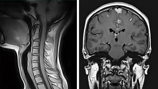
Magnetic resonance imaging (MRI), also known as magnetic resonance tomography (MRT) or simply RM, works on a completely different principle than X-rays and does not use radiation.
The term "tomography" simply means "layered imaging". While RMT includes the word tomography, magnetic resonance tomography should not be confused with computed tomography (CT).
The two techniques are fundamentally different, as distinct as day and night.
How does MRI work? In simple terms, during Magnetic Resonance Imaging (MRI), the body is exposed to a powerful rotating magnetic field. The water molecules contained in soft tissues are induced to vibrate at their specific resonance frequency, aligning themselves like magnets.
When the magnetic impulse stops, depending on the type of tissue and its specific water content, the molecules return to their initial state at different rates, allowing the differentiation of body tissues in the acquired image. The image acquisition occurs layer by layer and takes a relatively long time, up to 30-40 minutes, depending on the area to be scanned.
While an X-ray or Computed Tomography (CT) provides an excellent view of bones at the expense of other tissues, an MRI allows better visualization of soft tissues rather than bones. An MRI, like an X-ray, only provides a two-dimensional view of the scanned area. These two factors make MRI unsuitable for correctly identifying the position of the Atlas in three spatial planes. The major advantage of MRI is that it produces images without using harmful ionizing radiation, but unfortunately, it only offers a partial view of what is relevant to us.
Computed Tomography – 3D Spiral CT
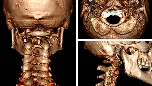
In simple terms, a Computed Tomography or CT, is an imaging technique that captures hundreds of "X-rays" from multiple perspectives, which are then assembled into a single three-dimensional image using dedicated software. Modern machines use spiral technology, where the X-ray tube and the opposing sensor rotate continuously around the patient while the table on which they lie moves slowly. Early scanners were called Axial Computed Tomography (ACT) because they only allowed image acquisition on a single axis.
The downside of this type of exam is the large amount of radiation (X-rays) to which one is exposed. The radiation from a CT scan of the skull is equivalent to 250 chest X-rays or 2000 microsieverts.
CBCT – Cone Beam Computed Tomography
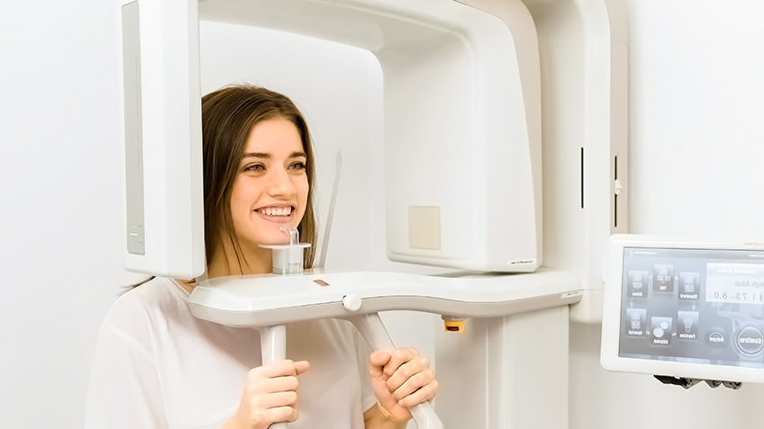
Cone Beam Computed Tomography (CBCT), known as Cone Beam CT, is primarily used in orthodontics. The main advantage of CBCT compared to a standard CT is undoubtedly the lower amount of radiation to which one is exposed.
Depending on the model of the scanner used, the absorbed radiation dose varies from 20 microsieverts in the latest-generation devices to 120-160 microsieverts in older devices. In the best-case scenario, the radiation is 100 times lower than that of a Spiral CT.
In terms of image quality, Spiral CT and Cone Beam CT are comparable, but Cone Beam CT has the advantage of producing fewer artifacts.
However, the lower intensity of the emitted radiation results in a reduced ability to distinguish bones from soft tissues, a limitation that can make it challenging to accurately visualize the Atlas. The Cone Beam CT, widely used in dentistry, is optimized to capture images of the jaw, which is covered by a thin layer of tissue, whereas the Atlas is located much deeper. This limitation can be overcome by increasing the radiation dose. Despite this, the absorbed radiation dose remains significantly lower compared to a spiral CT, making the CBCT a safer option in terms of radiation exposure.
The Cone Beam CT scan is performed in an upright position, without needing to lie down in a tube, a feature particularly appreciated by individuals suffering from claustrophobia. Another advantage is that the cervical vertebrae are under load, avoiding the flattening of the physiological curve of the spine, which inevitably occurs when lying down. This way, it is possible to determine not only vertebral alignment but also the presence of a possible cervical straightening.
Another factor to consider is the following: in dentistry, small-sized sensors, typically around 7 cm, are used. However, these dimensions are insufficient to capture the entire contour of the skull. To obtain adequate images, a sensor of at least 15 cm is required. As explained below, if the entire circumference of the skull is not captured, the images may become unusable for our purposes.
Therefore:
- A sensor larger than 15 cm is required.
- The radiation dose must be increased to better differentiate tissues.
It is undeniably more challenging to find a center properly equipped to perform a Cone Beam CT scan compared to one with a spiral CT scanner, as the latter is much more common. However, the effort required to locate the right center is certainly justified when it comes to protecting your health.
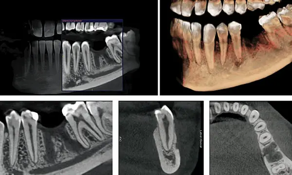
An additional advantage of a well-performed Cone Beam CT scan is the ability to visualize the roots of the teeth and the surrounding bone with a precision impossible to achieve with conventional or panoramic X-rays.
In two-dimensional images, the multiple roots of molar teeth often overlap, obscuring one another. With a CBCT, however, your dentist will be able to identify hidden infections that would otherwise remain undetected.
Oral cavity problems tend to increase with age and can cause serious chronic health issues. Take advantage of this opportunity, especially if you already need to undergo a CBCT!
A CT Scan Should Only Be Performed in Specific Circumstances
A CT scan does not provide information about muscle tension or joint functionality. For this reason, regardless of the availability of a CT scan, the individual must still be manually and expertly evaluated by an Atlantomed specialist.
The CT scan should be considered as a support for complex cases rather than a first-line investigation. You will understand that it is neither logical nor effective to undergo a CT scan on your own initiative in the hope of avoiding a trip. You may have a relatively well-aligned Atlas, but cervical musculature so contracted and dysfunctional that a treatment with AtlantoVib would still be necessary and beneficial. In the Atlantomed approach, unlike conventional medicine, the simplest, most sensible, and least invasive path is prioritized, which proves effective in 90% of cases. Only in the remaining 10% are more complex solutions resorted to when necessary. This approach minimizes unnecessary exams and treatments.
The rotation of the Atlas or Axis is only one of the factors assessed as an indication for treatment. Therefore, you will understand that it makes no sense to expose yourself to radiation without first being carefully examined by an Atlas specialist. In most cases, a CT scan is not needed and should only be considered at later stages, if strictly necessary. If this exam has already been conducted in the past, it is important for the therapist to analyze it before the session. For this, volumetric CT data is required, not just the medical report.
How to Properly Perform a CT Scan to Visualize Atlas Misalignment
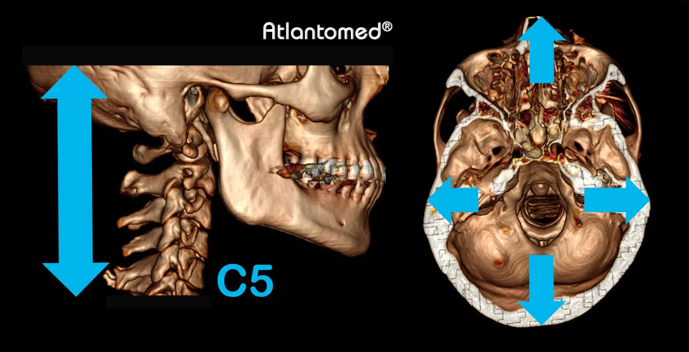
To properly evaluate the upper cervical region, it is necessary to acquire images of the correct anatomical area. The ideal portion to visualize should include the lower third of the skull and extend to the C5-C6 vertebrae, provided the sensor size allows it.
It is not necessary to irradiate the entire skull upward, nor to extend the scan too far down the cervical spine. Irradiating a larger anatomical portion than needed is not only unnecessary for the study’s purpose but also exposes the body to unwarranted radiation. Scanning below C5-C6 would unnecessarily irradiate the thyroid, an organ particularly sensitive to radiation.
It is crucial that the entire circumference of the skull is visible in the images. Missing any part, whether frontal (including the teeth), posterior, or lateral, can deprive the specialist of essential references for an adequate evaluation, potentially rendering the images unusable. Proper image acquisition is therefore essential for accurate assessment.
How to Properly Evaluate a CT Scan?
There are certain circumstances that require a CT scan evaluation by an Atlas specialist, perhaps because a scan was done previously, a new one was performed after unsuccessful treatment, or in special cases where malformations or contraindications are present.
Evaluating vertebral alignment via a CT scan begins with reconstructing the volumetric data into a 3D image. This allows the skull to be observed and analyzed from different angles, eliminating sections that might obscure relevant details. This requires the appropriate software, proper knowledge, a clear understanding of what to look for, and focus. Therefore, you will understand that it is absolutely useless to show us a single image extracted from your CD, as often happens, regardless of what that image may be! Without the listed elements, the analysis becomes a pointless and misleading exercise. If we do not receive the file with the data but only individual images, we cannot derive anything from it!
To perform 3D reconstruction and adequately evaluate the cervical region, all volumetric CT data is required, that is, the folder containing the images in DICOM format, approximately 100-500MB, found on the CD. All other files are superfluous and should not be sent. The folder in question is usually named DICOM but could have a different name assigned by the radiologist. If the folder is less than 50MB, you are NOT sending the CORRECT one! As mentioned earlier, it is useless to send X-rays, MRI scans, medical or radiological reports unless explicitly requested for specific reasons. If you have read and understood what has been explained and wish to send us your CT images, the easiest, fastest, and most effective way is to send only the folder containing the volumetric data using the wetransfer.com service.
How Can I Tell If My Atlas Is Misaligned?
The radiologist's CD includes a basic utility program that allows you to view the images layer by layer in 2D across three different planes. It may seem obvious to identify an Atlas misalignment, but what you think you see in 2D mode is only accurate under very specific conditions.
To confirm a misalignment in 2D visualization, some preliminary adjustments are necessary. It is impossible to correctly assess the position of the Atlas in relation to the skull if the latter is not first aligned on its three planes. During a scan, the head is rarely in a perfectly orthogonal position, an aspect that is often overlooked but must be considered and corrected before analyzing the images, especially in 2D visualization. The question to ask is: what is misaligned relative to what? If, from the start, the entire head appears unaligned and not orthogonal in the scan you are observing, any internal structure you examine will be similarly affected. Furthermore, if the CT scan lacks the complete contour of the skull, properly aligning the image becomes impossible, and what you believe you are seeing may not correspond to reality. In other words, without stable references to verify whether the entire head is tilted or not, how can you expect to understand what is misaligned internally? Yet, many make diagnoses in this absurd manner, without truly understanding what they are observing. There are also other significant challenges to consider, but these are not elaborated here for brevity.
These adjustments are systematically ignored, and if the CT scans are additionally done "poorly," as often happens, they become impossible to apply! Looking at a single layer image from a CT scan, without first ensuring that the skull has been accurately aligned on its axes, renders any analysis meaningless.
With 3D reconstruction of the images, it is possible to overcome some deficiencies in the original acquisition, making the evaluation of misalignments much easier and more straightforward. However, it remains necessary to rotate the 3D image of the skull multiple times across various axes to observe the details useful for assessing misalignment.
IMPORTANT: as mentioned at the beginning and reiterated here to emphasize its importance, a radiologist is trained to identify specific structural alterations and defined pathologies, but Atlas misalignment is not among the issues that are commonly looked for. Consequently, it is unlikely that a radiologist can provide an accurate evaluation of this aspect. It is very rare to find indications of vertebral misalignments in radiological reports, and even when they are present, they may be imprecise or incorrect. We emphasize this point because many people believe their spine is perfectly aligned simply because nothing specific is mentioned in the report. The phrase "the doctor said everything is fine" is something we hear often, but unfortunately, it may not reflect reality. For this reason, we independently examine and evaluate the CT images, regardless of the reports. We are committed to analyzing the images in detail and sharing with you everything we find, including any anomalies or misalignments that may have been overlooked in a standard radiological evaluation.
Effects on the Axis and Other Cervical Vertebrae
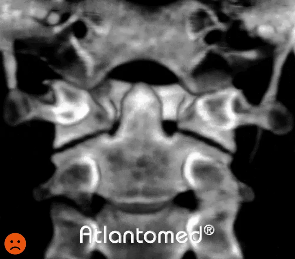
BEFORE
AFTER ATLANTOMED
This three-dimensional reconstruction, obtained using a spiral CT scan, provides a detailed view of the upper cervical vertebrae, observed from behind, both before and after the correction of the Atlas vertebra using the Atlantomed vibro-resonance technique.
The post-treatment image clearly shows a significant improvement in the alignment of the first Atlas vertebra relative to the occiput and the second cervical vertebra (Axis). This new balance translates into a more natural and symmetrical arrangement of the upper cervical structures.
Specifically, the Axis odontoid process appears noticeably more centered in its articulation with the Atlas, improving the distribution of mechanical forces throughout the cervical region. Additionally, the skull, which before treatment appeared tilted to the right, has regained a more balanced position, indicating a restoration of structural harmony between the skull and the spine.
Conclusions
Once the differences between various imaging diagnostic methods are understood, it is essential to make some important considerations: ionizing radiation, such as X-rays, is not beneficial to health and should be used sparingly, only when absolutely necessary and when the risk-benefit ratio clearly favors the benefit.
Exposure to radiation increases the risk of developing cancers and leukemias. For this reason, today in many contexts, MRI is preferred over CT scans, provided the clinical circumstances allow. However, as extensively explained earlier, MRI is not suitable for evaluating the Atlas.
If manual evaluation of the vertebrae proves insufficient, and a CT scan allows for a targeted and effective correction resulting in the disappearance of symptoms, then the radiological examination is justified.
However, it would not be justified if the same result could have been achieved solely through the manual examination performed by an Atlantomed specialist. This means that the correct approach is to first visit an Atlantomed Center to determine the best course of action together. If, out of convenience or to avoid travel, you decide to have a CT scan performed in advance, be aware that you are not acting responsibly to preserve your long-term health. On the other hand, if you already have this test done previously, it is advisable to bring it with you or send it in advance. We refer exclusively to spiral CT or Cone Beam CT (CBCT), not MRI. If you do not understand the difference, we invite you to reread the explanations carefully.
Many people undergo CT scans with a certain nonchalance, almost as if it were a routine action, without considering the potential long-term consequences of radiation exposure. Often, the attending radiologist does not have the time or opportunity to provide adequate information at the time of the exam. For this reason, we have explored the topic in detail, so you can make an informed and sensible decision.
A preliminary CT scan is NOT required for Atlas correction treatment. Only in specific cases, if the Atlantomed specialist encounters difficulty determining the Atlas position through manual examination, can it be evaluated together how to proceed.
Important Notice
It is important to clarify that the Atlantomed specialist examines the images exclusively to evaluate the position of the vertebrae to determine how to proceed with the correction. This is not a medical diagnosis and should not be interpreted as such. Atlas misalignment is not a disease.
If you seek a diagnosis of the pathological condition of your spine or the state of your dental roots, we recommend consulting a competent radiologist or dentist.
It is not our role to receive your MRI or medical reports to provide a diagnosis. Atlantomed is NOT a medical treatment, but a purely mechanical treatment; for this reason, the Atlas specialist does not intend to replace your trusted physician in any way.
Read this page carefully: Who is Atlantomed For?
What people say about us
The only ones with 10,000 testimonials and reviews in several languages! Click to access the related platforms and read many opinions, feedbacks, experiences and testimonials after the Atlantomed vibro-resonance Atlas correction. Be wary of imitations.
Written by: Alfredo Lerro


